HyStem / Hyaluronic Acid
General Hyaluronic Acid Information
Hyaluronic acid, sometimes referred to a hyaluronan or HA, is the most abundant glycosaminoglycan in the body with being an important major component of several tissues throughout the body. While it is abundant in extracellular matrices, hyaluronan also contributes to tissue hydrodynamics, movement and proliferation of cells, and participates in a number of cell surface receptor interactions.
Hyaluronic acid is a polymer of disaccharides, themselves composed of D-glucuronic acid and N-acetyl-D-glucosamine, linked via alternating β-(1→4) and β-(1→3) glycosidic bonds. Hyaluronic acid can be 25,000 disaccharide repeats in length. Polymers of hyaluronic acid can range in size from 5,000 to 20,000,000 Da in vivo.
Rheological Properties of Hyaluronic Acid
As a polymer in solution, shows non-Newtonian and viscoelastic behavior. Non-Newtonian liquids exhibit viscosity dependence upon the applied shear conditions. The most common type of non-Newtonian behavior being shear-thinning, where viscosity decreases with increasing shear rate.
This characteristic is beneficial as it allows for the product to be manipulated and handled with greater ease, such as when pumped and filled in manufacturing processes or dispensed via a needle as occurs in extrusion 3D bioprinting. Once at rest, however the product regains its viscosity, helping maintain its position.
Hyaluronic acid is highly soluble and often exhibits very poor mechanical properties with rapid degradation behavior in vivo. Hyaluronic acid has been chemically and crosslinker-modified to improve its properties, including mechanical properties, viscosity, solubility, degradation, and biologic properties.
The chemical structure of hyaluronic acid can be covalently modified where the three most commonly used sites includes the carboxylic groups, hydroxyl group, and –NHCOCH3 groups.
Methacrylated Hyaluronic Acid (PhotoHA®)
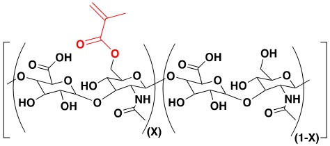
PhotoHA® is a methacrylated hyaluronic acid that can be photocrosslinked in the presence of a photoinitiator and light. Tunability is accomplished by altering the HA concentration, photocrosslinking time and intensity, or by adjusting the photoinitiator concentration.
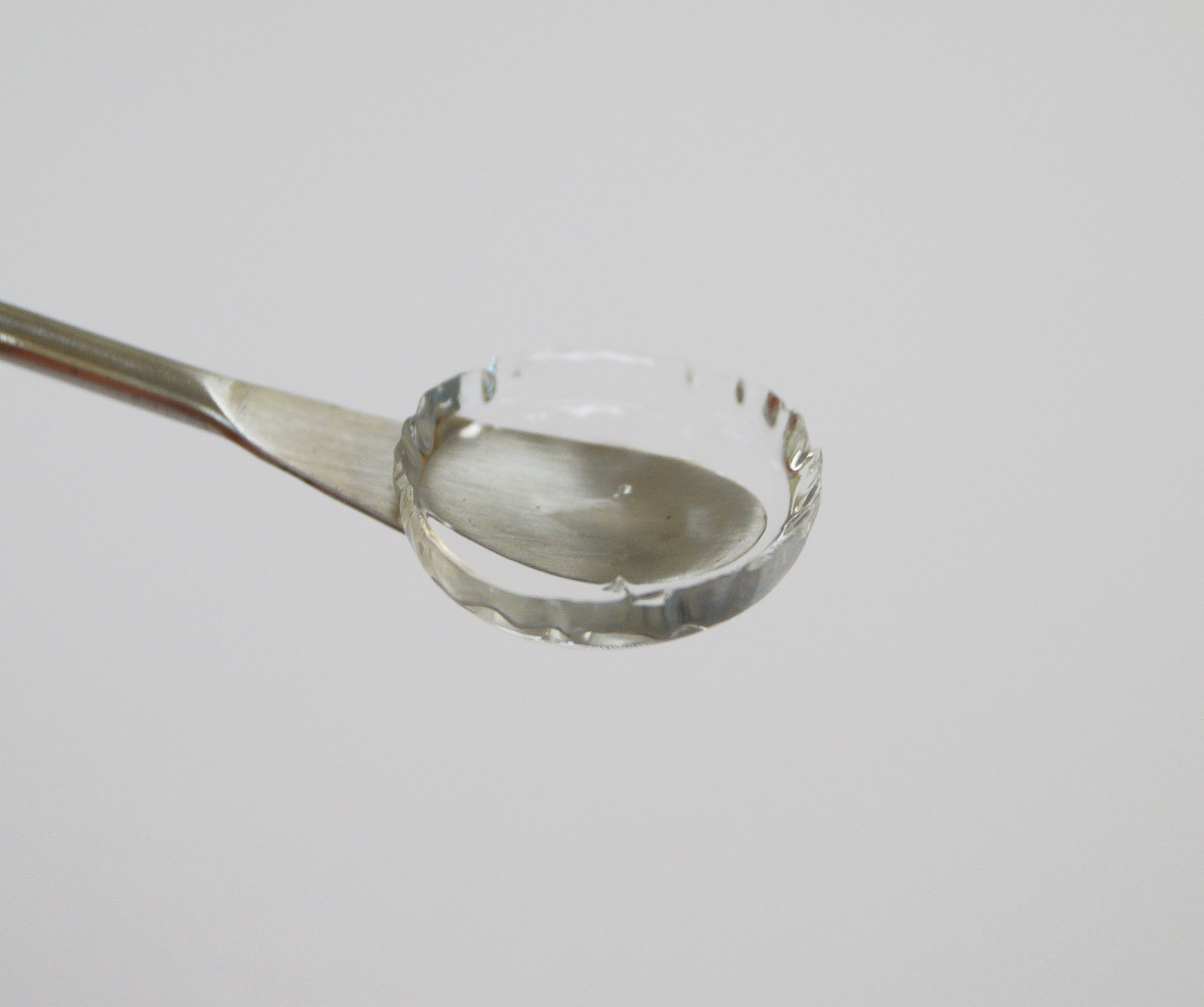
Crosslinked PhotoHA® hydrogels are strong and optically clear (as seen in the image above).
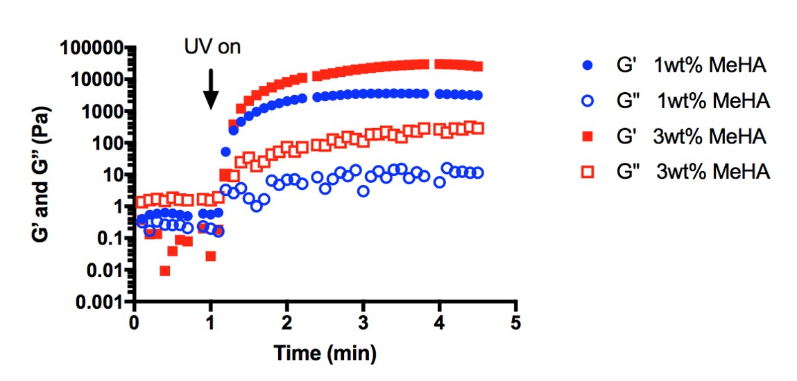
Rheological testing using Irgacure and UV light (365 nm) demonstrate a high rate of crosslinkability, tunability, and strength for PhotoHA® hydrogels (right).
3D Stem Cell Culture with HyStem Hydrogels
HyStem hydrogels are designed for stem cell culture. They are chemically defined and wholly animal free. They are based on hyaluronan (HA) – a component of the extracellular matrix (ECM) that is abundant in embryos and stem cell niches. HyStem hydrogels provide a compliant, viscoelastic matrix that is physiologically relevant for stem cells.
They emulate a stripped down version of the ECM to which additional components (such as growth factors and ECM proteins) can be added to recreate the composition of a specific niche.
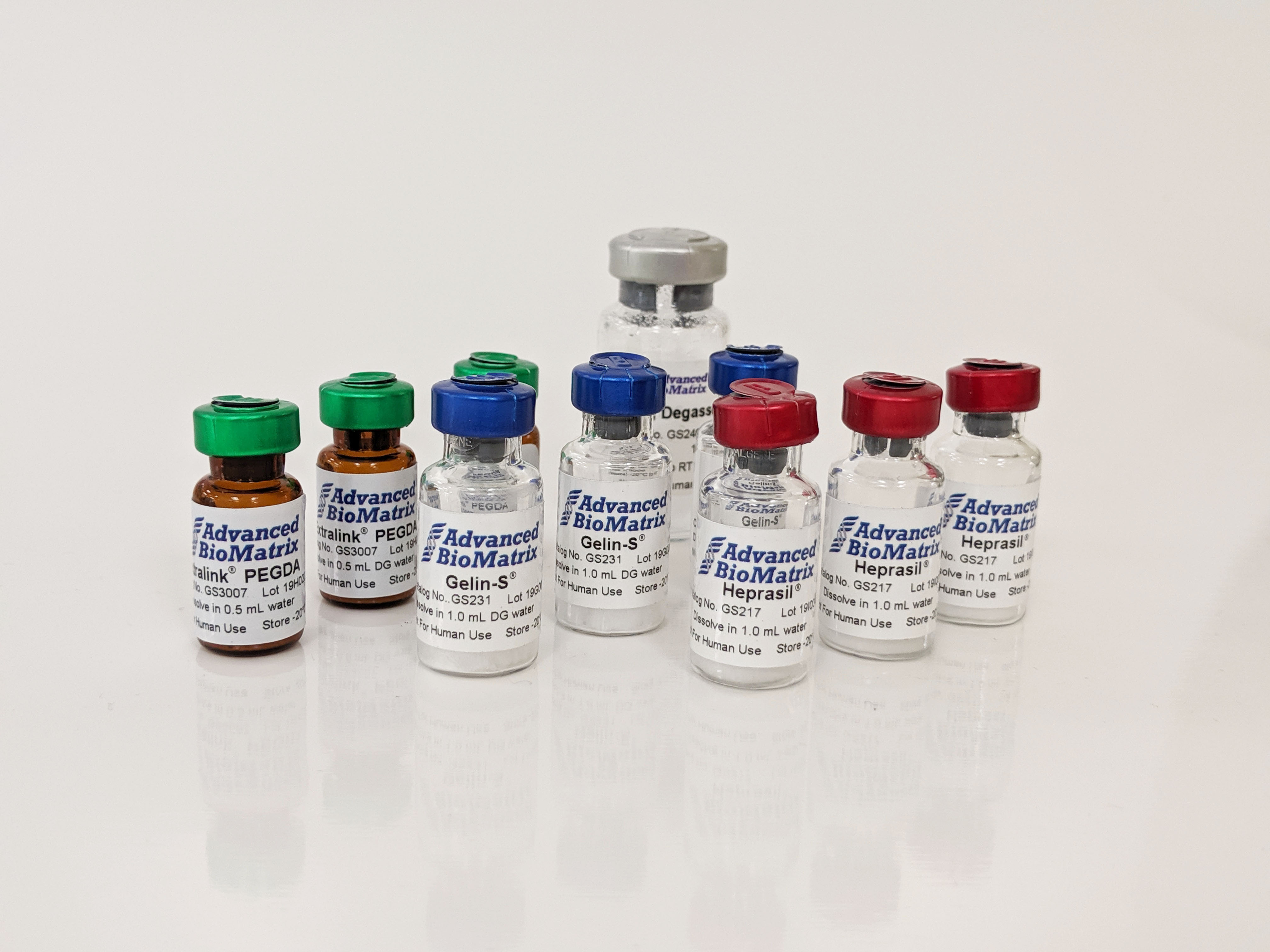
Three options for Stem Cell Culture:
- HyStem: Applications requiring attachment factor optimization. Extracellular matrix proteins can be mixed into the hydrogel and incorporated non-covalently before gelation. Alternatively, attachment peptides having an N-terminal cysteine can also be covalently linked to the matrix.
- HyStem-C: A general starting point for optimization of a cell's microenvironment. HyStem-C contains Gelin-S or thiolated gelatin (denatured collagen) which allows attachment of a wide variety of cell types and takes the guesswork out of the appropriate attachment factors to use.
- HyStem-HP: Applications requiring slow release of growth factors in a cell's microenvironment. HyStem-HP contains small amounts of thiolated heparin which ionically binds a wide variety of growth factors and slowly releases them over time.
Hyaluronan relevancy for stem cells
HyStem hydrogels are composed of HyStem (thiol-modified hyaluronan, HA) and Extralink (thiol-reactive crosslinking agent). HA is the simplest glycosaminoglycan (a class of negatively charged polysaccharides) and a major constituent of the ECM. Embryonic ECM possesses a high quantity of glycosaminoglycans of which HA is predominant. Human embryonic stem cells (H1, H9 and H13 lines) have been shown to express both CD44 and CD168 (RHAMM), which are HA receptors. Human embryonic stem cells (hESCs) have also been cultivated by encapsulating them in HA hydrogels. These cells were grown for 15 days without detectable differentiation. After recovery from the hydrogels, the hESCs could be differentiated using endothelial growth media supplemented with VEGF.
HA is also present in large amounts in the ECM of embryonic livers, fetal livers and the putative stem cell niche within the liver (the Canals of Hering). Hepatic stem and progenitors cells (hepatoblasts) have been successfully cultured for over 4 weeks without differentiation when encapsulated in HA hydrogels and grown with a defined media (Kubota’s medium). Additionally, these hepatic progenitor cells express CD44 at high levels.
