-
Collagen
-
Type I - Atelocollagen
- PureCol® Solution, 3 mg/ml (bovine) #5005
- Nutragen® Solution, 6 mg/ml (bovine) #5010
- FibriCol® Solution, 10 mg/ml (bovine) #5133
- PureCol® EZ Gel, Solution, 5 mg/ml (bovine) #5074
- PureCol® Lyophilized, 15 mg (bovine) #5006
- VitroCol® Solution, 3 mg/ml (human) #5007
- VitroCol® Lyophilized, 15 mg (human) #5008
-
Type I - Telocollagen
- TeloCol®-3 Solution, 3 mg/ml (bovine) #5026
- TeloCol®-6 Solution, 6 mg/ml (bovine) #5225
- TeloCol®-10 Solution, 10 mg/ml (bovine) #5226
- RatCol™ for 2D and 3D, Solution, 4 mg/ml (rat) #5153
- RatCol™ High Concentration, Solution, 10 mg/ml (rat)
- RatCol™ lyophilized, 100 mg (rat)
- RatCol™ for Coatings, Solution, 4 mg/ml (rat) #5056
- Type I - Insoluble Collagen
- Type I - Bioinks
- Type II Collagen
- Type III Collagen
- Type IV Collagen
- Collagen Standard
-
PureCol® Collagen Coated Plates
- Collagen Coated T-25 Flasks #5029
- Collagen Coated 6-well Plates #5073
- Collagen Coated 12-well Plates #5439
- Collagen Coated 24-well Plates #5440
- Collagen Coated 48-well Plates #5181
- Collagen Coated 96-well Plates #5072
- Collagen Coated 384-well Plates #5380-5EA
- Collagen Coated 100 x 20 mm Dishes #5028
- MatTek Glass-Bottom Dishes
- MatTek Multi-Well Plates
- Collagen Scaffolds
- Collagen Hybridizing Peptides
-
Type I - Atelocollagen
- Tunable Stiffness
- CytoSoft™ Rigidity Plates
-
Bioprinting
- Support Slurry for FRESH Bioprinting
-
Bioinks for Extrusion Bioprinting
- Lifeink® 200 Collagen Bioink (35 mg/ml) #5278
- Lifeink® 220 Collagen Bioink (70 mg/ml) #5343
- Lifeink® 240 Acidic Collagen Bioink (35 mg/ml) #5267
- Lifeink® 260 Acidic Collagen Bioink (70 mg/ml) #5358
- GelMA Bioink
- GelMA A Bioink
- GelMA C Bioink
- Pluronic F-127 40% Sterile Solution
- GelMA 20% Sterile Solution
- Alginate 5% Sterile Solution
- Photoinitiators
- Bioinks for BIONOVA X
- Bioinks for Lumen X
- DLP Printing Consumables
-
Create Your Own Bioinks
- PhotoCol® Methacrylated Collagen
- PhotoGel® Methacrylated Gelatin 95% DS
- PhotoGel® Methacrylated Gelatin 50% DS
- PhotoHA®-Stiff Methacrylated Hyaluronic Acid
- PhotoHA®-Soft Methacrylated Hyaluronic Acid
- PhotoAlginate® Methacrylated Alginate
- PhotoDextran® Methacrylated Dextran
- PEGDA (Various Molecular Weights)
- Silk Fibroin, Solution
- PhotoSericin® Methacrylated Sericin
- Bioprinters
-
3D Hydrogels
- Thermoreversible Hydrogel
- Silk Fibroin
-
Type I Collagen for 3D Hydrogels
- PureCol® Solution, 3 mg/ml (bovine) #5005
- Nutragen® Solution, 6 mg/ml (bovine) #5010
- FibriCol® Solution, 10 mg/ml (bovine) #5133
- PureCol® EZ Gel, Solution, 5 mg/ml (bovine) #5074
- VitroCol® Solution, 3 mg/ml (human) #5007
- TeloCol®-3 Solution, 3 mg/ml (bovine) #5026
- TeloCol®-6 Solution, 6 mg/ml (bovine) #5225
- TeloCol®-10 Solution, 10 mg/ml (bovine) #5226
- RatCol® for 3D gels, Solution, 4 mg/ml (rat) #5153
- HyStem® Thiolated Hyaluronic Acid
- Methacrylated Collagen
- Methacrylated Gelatin
- Methacrylated Hyaluronic Acid
- Diacrylates
- Collagen Sponges
- Methacrylated Polysaccharides
- Spheroids and Organoids
- Extracellular Matrices
- HyStem / Hyaluronic Acid
-
Adhesion Peptides / Proteins
-
Recombinant Adhesion Proteins
- CD2, 0.5 mg/ml #5086
- CDH3, 0.5 mg/ml #5124
- CDH13, 0.5 mg/ml #5125
- CD14, 0.5 mg/ml #5089
- CDH18, 0.5 mg/ml #5090
- CD40, 0.5 mg/ml #5093
- CD86, 0.5 mg/ml #5096
- CD164, 0.5 mg/ml #5100
- CD270, 0.5 mg/ml #5127
- CD274, 0.5 mg/ml #5126
- CD276, 0.5 mg/ml #5123
- E-Cadherin (CD324), 0.5 mg/ml #5085
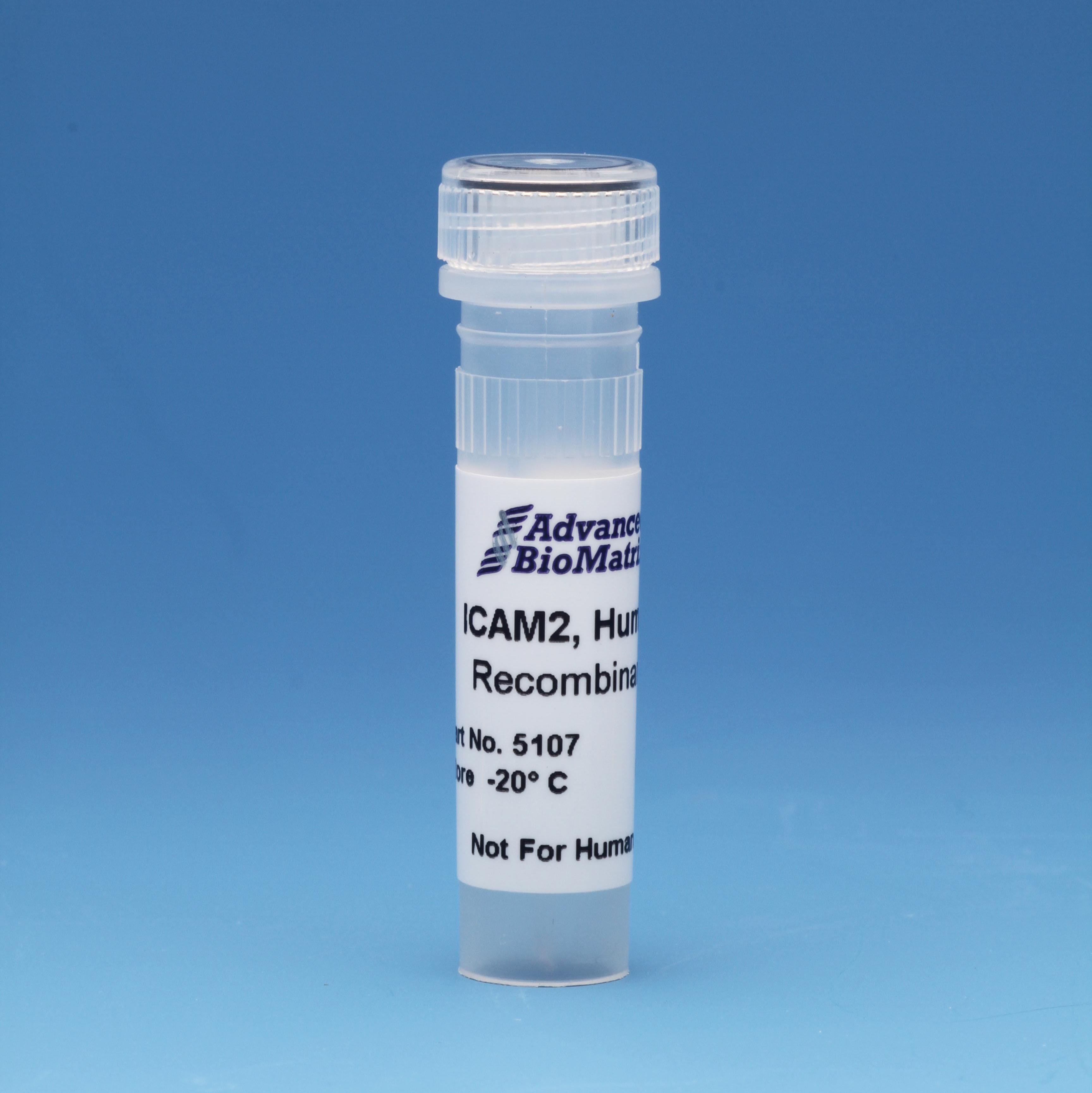
- ICAM2, 0.5 mg/ml #5107
- Adhesion Peptides
- Collagen Hybridizing Peptides
-
Recombinant Adhesion Proteins
- Reagents
- Assays
ICAM2
Solution, 0.5 mg/ml (Recombinant)
Catalog #5107
ICAM2
Solution, 0.5 mg/ml (Recombinant)
Catalog #5107
The protein encoded by human ICAM2 gene is a member of the intercellular adhesion molecule (ICAM) family. All ICAM proteins are type I transmembrane glycoproteins, contain 2-9 immunoglobulin-like C2-type domains, and bind to the leukocyte adhesion LFA-1 protein.
Product Description
The protein encoded by human ICAM2 gene is a member of the intercellular adhesion molecule (ICAM) family. All ICAM proteins are type I transmembrane glycoproteins, contain 2-9 immunoglobulin-like C2-type domains, and bind to the leukocyte adhesion LFA-1 protein. This protein may play a role in lymphocyte recirculation by blocking LFA-1-dependent cell adhesion. It mediates adhesive interactions important for antigen-specific immune response, NK-cell mediated clearance, lymphocyte recirculation, and other cellular interactions important for immune response and surveillance. Several transcript variants encoding the same protein have been found for this gene.
Full-length extracellular domain of human ICAM2 gene (25-223 aa) was constructed with 29 N-terminal T7/His tag and expressed in E. coli as inclusion bodies.
The final product was refolded using a unique “temperature shift inclusion body refolding” technology and chromatographically purified.
| Parameter, Testing, and Method | ICAM2 #5107 |
| Quantity | 0.1 mg |
| Volume | 0.2 mL |
| Concentration | 0.5 mg/mL |
| Purity - SDS PAGE Electrophoresis | > 90% |
| Formulation | Formulated in 20 mM pH 8.0 TRIS-HCL Buffer, with proprietary formulation of NaCl, KCl, EDTA, L-Arginine, DTT and Glycerol |
| Form | Solution |
| Production Type | Recombinant - E. Coli |
| Storage Temperature | -20°C |
| Shelf Life | Minimum of 6 months from date of receipt |
| Sterilization Method | Filtration |
| Cell Assay | Pass |
| Sterility - USP modified | No Growth |
| Accession Number | NP_000864 |
| Recombinant Protein Sequence | MASMTGGQQMGRGHHHHHHGNLYFQGGEFELKVFEVHVRPKKLAVEPKGSLEV NCSTTCNQPEVGGLETSLDKILLDEQAQWKHYLVSNISHDTVLQCHFTCSGKQ ESMNSNVSVYQPPRQVILTLQPTLVAVGKSFTIECRVPTVEPLDSLTLFLFRGNET LHYETFGKAAPAPQEATATFNSTADREDGHRNFSCLAVLDLMSRGGNIFHKHSA PKMLEIYEPVSDSQ |
Directions for Use
Download the full PDF version or continue reading below:
Use these recommendations as guidelines to determine the optimal coating conditions for your culture system.
- Thaw ICAM2 and dilute to desired concentration using serum-free medium or PBS. The final solution should be sufficiently dilute so that the volume added covers the surface evenly. Note: Use 1 ml PBS per well in a 6-well plate.
- Add 1 – 10 µg protein to each well and incubate at 2 to 10°C overnight.
- After incubation, aspirate remaining material.
- Plates are ready for use. They may also be stored at 2-8°C damp or air dried if sterility is maintained.
Coating this recombinant protein at 1-10 ug / well (6 well plate) in neuronal cell specific medium can be used for 1) human lymphocyte cell / receptor interaction study in vitro and 2) as a culture matrix protein for anti-tumor immuno-response study in vitro.
Product References
References for Lifeink® collagen bioinks:
Lan, X. et al. In vitro maturation and in vivo stability of bioprinted human nasal cartilage. Journal of Tissue Engineering 13, 204173142210863 (2022).
Stocco, T. D., Moreira Silva, M. C., Corat, M. A., Gonçalves Lima, G. & Lobo, A. O. Towards bioinspired meniscus-regenerative scaffolds: Engineering A novel 3D bioprinted patient-specific construct reinforced by biomimetically aligned nanofibers. International Journal of Nanomedicine Volume 17, 1111–1124 (2022).
Lee, A. et al. 3D bioprinting of collagen to rebuild components of the human heart. Science 365, 482–487 (2019).
Maxson, Eva L., et al. "In vivo remodeling of a 3D-Bioprinted tissue engineered heart valve scaffold." Bioprinting (2019): e00059.
Filardo, G. et al.Patient-specific meniscus prototype based on 3D bioprinting of human cell-laden scaffold. Bone & Joint Research 8,101–106 (2019).
Schmitt, T. Analysis and Classification of 3-D Printed Collagen-Bioglass Matrices for Cellular Growth Utilizing Artificial Neural Networks. University Thesis (2018).
Balakhovsky, Y. M., Ostrovskiy, A. Y. & Khesuani, Y. D. Emerging Business Models Toward Commercialization of Bioprinting Technology. 3D Printing and Biofabrication1–22 (2017). doi:10.1007/978-3-319-40498-1_25-1
Fox, S. et al. A simplified fabrication technique for cellularized high-collagen dermal equivalents. Biomedical Materials14,041001 (2019).
Lin, H.-H., Chao, P.-H. G., Tai, W.-C. & Chang, P.-C. 3D-printed collagen-based waveform microfibrous scaffold for periodontal ligament reconstruction. International Journal of Molecular Sciences 22, 7725 (2021).
Engberg, A., Stelzl, C., Eriksson, O., O’Callaghan, P. & Kreuger, J. An open source extrusion bioprinter based on the E3D motion system and Tool Changer to enable fresh and multimaterial bioprinting. Scientific Reports 11, (2021).
Tashman, J., et al. "In Situ Volumetric Imaging and Analysis of FRESH 3D Bioprinted Constructs Using Optical Coherence Tomography." BioRxiv. (2021)
Product Certificate of Analysis
No result for .
Product Disclaimer
This product is for R&D use only and is not intended for human or other uses. Please consult the Material Safety Data Sheet for information regarding hazards and safe handling practices.


