-
Collagen
-
Type I - Atelocollagen
- PureCol® Solution, 3 mg/ml (bovine) #5005
- Nutragen® Solution, 6 mg/ml (bovine) #5010
- FibriCol® Solution, 10 mg/ml (bovine) #5133
- PureCol® EZ Gel, Solution, 5 mg/ml (bovine) #5074
- PureCol® Lyophilized, 15 mg (bovine) #5006
- VitroCol® Solution, 3 mg/ml (human) #5007
- VitroCol® Lyophilized, 15 mg (human) #5008
-
Type I - Telocollagen
- TeloCol®-3 Solution, 3 mg/ml (bovine) #5026
- TeloCol®-6 Solution, 6 mg/ml (bovine) #5225
- TeloCol®-10 Solution, 10 mg/ml (bovine) #5226
- RatCol™ for 2D and 3D, Solution, 4 mg/ml (rat) #5153
- RatCol™ High Concentration, Solution, 10 mg/ml (rat)
- RatCol™ lyophilized, 100 mg (rat)
- RatCol™ for Coatings, Solution, 4 mg/ml (rat) #5056
- Type I - Insoluble Collagen
- Type I - Bioinks
- Type II Collagen
- Type III Collagen
- Type IV Collagen
- Collagen Standard
-
PureCol® Collagen Coated Plates
- Collagen Coated T-25 Flasks #5029
- Collagen Coated 6-well Plates #5073
- Collagen Coated 12-well Plates #5439
- Collagen Coated 24-well Plates #5440
- Collagen Coated 48-well Plates #5181
- Collagen Coated 96-well Plates #5072
- Collagen Coated 384-well Plates #5380-5EA
- Collagen Coated 100 x 20 mm Dishes #5028
- MatTek Glass-Bottom Dishes
- MatTek Multi-Well Plates
- Collagen Scaffolds
- Collagen Hybridizing Peptides
-
Type I - Atelocollagen
- Tunable Stiffness
- CytoSoft™ Rigidity Plates
-
Bioprinting
- Support Slurry for FRESH Bioprinting
-
Bioinks for Extrusion Bioprinting
- Lifeink® 200 Collagen Bioink (35 mg/ml) #5278
- Lifeink® 220 Collagen Bioink (70 mg/ml) #5343
- Lifeink® 240 Acidic Collagen Bioink (35 mg/ml) #5267
- Lifeink® 260 Acidic Collagen Bioink (70 mg/ml) #5358
- GelMA Bioink
- GelMA A Bioink
- GelMA C Bioink
- Pluronic F-127 40% Sterile Solution
- GelMA 20% Sterile Solution
- Alginate 5% Sterile Solution
- Photoinitiators
- Bioinks for BIONOVA X
- Bioinks for Lumen X
- DLP Printing Consumables
-
Create Your Own Bioinks
- PhotoCol® Methacrylated Collagen
- PhotoGel® Methacrylated Gelatin 95% DS
- PhotoGel® Methacrylated Gelatin 50% DS
- PhotoHA®-Stiff Methacrylated Hyaluronic Acid
- PhotoHA®-Soft Methacrylated Hyaluronic Acid
- PhotoAlginate® Methacrylated Alginate
- PhotoDextran® Methacrylated Dextran
- PEGDA (Various Molecular Weights)
- Silk Fibroin, Solution
- PhotoSericin® Methacrylated Sericin
- Bioprinters
-
3D Hydrogels
- Thermoreversible Hydrogel
- Silk Fibroin
-
Type I Collagen for 3D Hydrogels
- PureCol® Solution, 3 mg/ml (bovine) #5005
- Nutragen® Solution, 6 mg/ml (bovine) #5010
- FibriCol® Solution, 10 mg/ml (bovine) #5133
- PureCol® EZ Gel, Solution, 5 mg/ml (bovine) #5074
- VitroCol® Solution, 3 mg/ml (human) #5007
- TeloCol®-3 Solution, 3 mg/ml (bovine) #5026
- TeloCol®-6 Solution, 6 mg/ml (bovine) #5225
- TeloCol®-10 Solution, 10 mg/ml (bovine) #5226
- RatCol® for 3D gels, Solution, 4 mg/ml (rat) #5153
- HyStem® Thiolated Hyaluronic Acid
- Methacrylated Collagen
- Methacrylated Gelatin
- Methacrylated Hyaluronic Acid
- Diacrylates
- Collagen Sponges
- Methacrylated Polysaccharides
- Spheroids and Organoids
- Extracellular Matrices
- HyStem / Hyaluronic Acid
-
Adhesion Peptides / Proteins
-
Recombinant Adhesion Proteins
- CD2, 0.5 mg/ml #5086
- CDH3, 0.5 mg/ml #5124
- CDH13, 0.5 mg/ml #5125
- CD14, 0.5 mg/ml #5089
- CDH18, 0.5 mg/ml #5090
- CD40, 0.5 mg/ml #5093
- CD86, 0.5 mg/ml #5096
- CD164, 0.5 mg/ml #5100
- CD270, 0.5 mg/ml #5127
- CD274, 0.5 mg/ml #5126
- CD276, 0.5 mg/ml #5123
- E-Cadherin (CD324), 0.5 mg/ml #5085
- ICAM2, 0.5 mg/ml #5107
- Adhesion Peptides
- Collagen Hybridizing Peptides
-
Recombinant Adhesion Proteins
- Reagents
- Assays
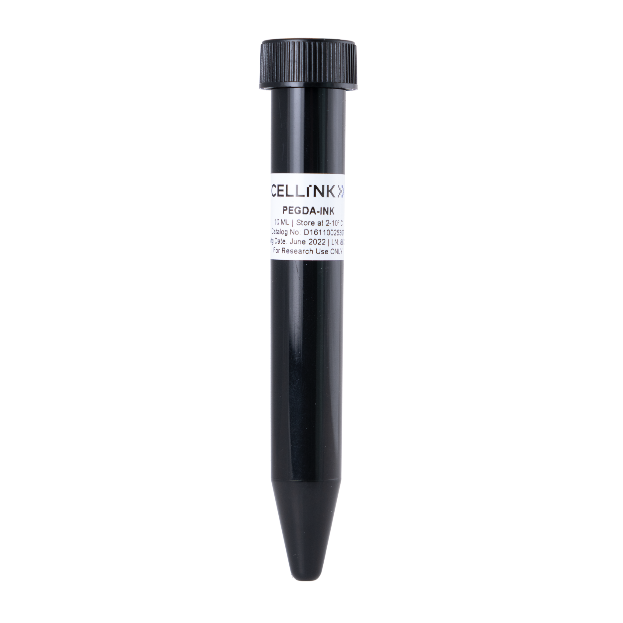
PEGDA-INK
Pre-Formulated Ink
Catalog #D16110025307
PEGDA-INK
Pre-Formulated Ink
Catalog #D16110025307
PEGDA-INK is 10mL of preformulated 50% PEGDA ink (700 MW) with 0.5% LAP that gels rapidly using the BIONOVA X DLP 3D Bioprinter.
Product Description
Polyethylene Glycol Diacrylate (PEGDA) hydrogels are powerful tools for uncovering basic cellular biology because they are considered biologically inert (“blank slate”) and their mechanical properties can be varied over a large range of moduli.
PEGDA is an emerging scaffold for tissue engineering and regenerative medicine since polymerization can occur rapidly at room temperature and requires low energy input, has high water content, is elastic, and can be customized to include a variety of biological molecules.
PEGDA is a crosslinker repeating polymer crosslinker and is >80% acrylated. It can be used as a thiol-reactive crosslinker in our HyStem systems at low concentration, or can be used to make a PEGDA-only hydrogel in the presence of a free-radical chain photoinitiator and light source. PEG-only gels do not typically support cell attachment without the incorporation of cellular attachment sites.
Applications
Regular and 3-D cell culture
Tissue engineering
Photolithography
Store PEGDA in the original vial unopened at -20°C for up to one year. Reconstituted PEGDA solutions can be stored at -20°C for ~ one month.
Product Applications
Read our HyStem eBrochure Here
or
Click on the title of the desired protocol to learn more:
2D Cell Growth on HyStem Hydrogels
HyStem 3D Cell Encapsulation for Cell Delivery Applications Guide
HyStem 3D Cell Encapsulation in hydrogels using 96-well plates
HyStem 3D Cell Encapsulation in hydrogels using TC Inserts
Enzyme Digestion of HyStem Hydrogels for Recovery of Encapsulated Cells
Fluorescent Labeling of HyStem Hydrogels
Cell Recovery from Surface of HyStem Hydrogels
HyStem Gelation Time Variation
Product References
Salinas CN and Anseth KS, The enhancement of chondrogenic differentiation of human mesenchymal stem cells by enzymatically regulated RGD functionalities. Biomaterials. (2008) 29:2370-7.
Kloxin AM, et al.Photodegradable Hydrogels for Dynamic Tuning of Physical and Chemical Properties Science (2009) 324, 59.
DeForest CA, Polizzotti BD, and Anseth KS, Sequential click reactions for synthesizing and patterning three-dimensional cell microenvironments Nat. Mater. (2009) 8: pp. 659-664.
Baird IS, Yau AY, and Mann BK, Mammalian cell-seeded hydrogel microarrays printed via dip-pin technology BioTech. (2008) 44:249-256.
Baek TJ et al, Photolithographic Fabrication of Poly(Ethylene Glycol) Microstructures for Hydrogel-based Microreactors and Spatially Addressed Microarrays J. Microbiol. Biotechnol. (2007) 17: 1826-1832.
Taite LJ, Rowland ML, Ruffino KA, Smith BR, Lawrence MB, West JL. Bioactive hydrogel substrates: probing leukocyte receptor-ligand interactions in parallel plate flow chamber studies. Ann Biomed Eng. (2006) 34:1705-11.
Du Y, Lo E, Ali S, Khademhosseini A. Directed assembly of cell-laden microgels for fabrication of 3D tissue constructs. Proc Natl Acad Sci U S A. (2008) 105:9522-7.
Khademhosseini A, Yeh J, Jon S, Eng G, Suh KY, Burdick JA, Langer R. Molded polyethylene glycol microstructures for capturing cells within microfluidic channels. Lab Chip. (2004) 4:425-30.
Liao H, Munoz-Pinto D, Qu X, Hou Y, Grunlan MA, Hahn MS. Influence of hydrogel mechanical properties and mesh size on vocal fold fibroblast extracellular matrix production and phenotype. Acta Biomater. (2008) 4:1161-71.
Patel PN, Smith CK, Patrick CW Jr. Rheological and recovery properties of poly(ethylene glycol) diacrylate hydrogels and human adipose tissue.J Biomed Mater Res A. (2005) 73:313-9.
Panda P et al, Stop-Flow Lithography to Generate Cell-Laden Microgel Particles. Lab Chip (2008) 8:1056-1061.
Ma PX and Elisseeff J editors, Scaffolds in Tissue Engineering, CRC Press Boca Raton, FL 2006.
Product Certificate of Analysis
No result for .
Product Disclaimer
This product is for R&D use only and is not intended for human or other uses. Please consult the Material Safety Data Sheet for information regarding hazards and safe handling practices.