High Concentration Rat Tail Collagen
Type I Rat Tail Collagen Solution, 10 mg/ml
Catalog #IKD119261001
High Concentration Rat Tail Collagen
Type I Rat Tail Collagen Solution, 10 mg/ml
Catalog #IKD119261001
Our high concentration rat tail collagen contains 10 mL of 10 mg/ml telocollagen. Being telocollagen, rat tail collagen will form firmer hydrogels than atelocollagen. Our collagen is highly purified, lot-to-lot consistent, and on-the-shelf.
Product Description
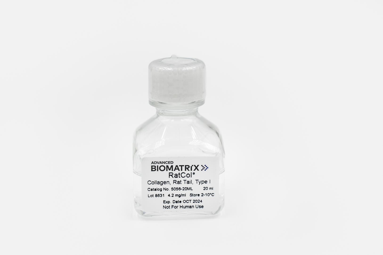
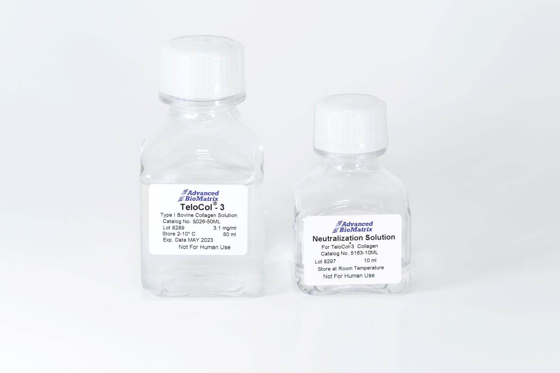
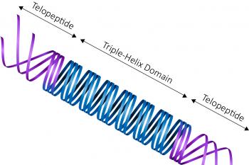
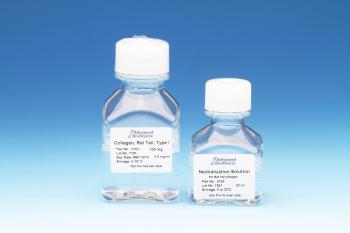
RatCol™ High Concentration Type I Acid Soluble Rat Tail Collagen contains 100 mg at a concentration of approximately 10 mg/mL in a 0.02M acetic acid solution (~pH 3). RatCol™ collagen is soluble telo-collagen. This collagen product is provided in user-friendly packaging for use and storage. This product is sterile filtered and is supplied as a ready to use solution.
This product is ideal for coating of surfaces, providing preparation of thin layers for culturing cells, or use as a solid gel. RatCol™ collagen is suitable for applications using a variety of cell lines including hepatocytes, fibroblasts and epithelial cells.
| Parameter, Testing, and Method | RatCol™ Type I Collagen #IKD119261001 |
| Sterilization Method | Filtration |
| Extraction Method | Acid - telocollagen |
| Form | Solution |
| Package Size | 100 mg (~10 mL) Kit |
| Storage Temperature of Collagen | 2-10°C |
| Storage Temperature of Neutralization Solution | Room Temperature |
| Shelf Life | Minimum of 6 months from date of receipt |
|
Collagen Concentration - Biuret |
~10.0 mg/mL |
|
Collagen Purity - Silver Staining |
>99% |
| pH | 3.0-3.8 |
| Kinetic Gel Test (Minutes) | <40 |
|
Gel Formation Tube Test (Minutes) |
<40 |
|
Electrophoretic Pattern - Coomassie Blue |
Characteristic |
| Sterility - USP modified | No growth |
| Endotoxin - LAL | <10.0 EU/mL |
| Osmolality (mOsmo H2O/kg) | <35 |
| Cell Attachment Assay | Pass |
| Source | Rat Tail Tendon |
| Hydrogel Young's Modulus E (Pa) | Characteristic |
Directions for Use
Recommended Volumes for 2D Coatings or 3D Hydrogels
Note - the 10 mg/ml collagen is highly viscous. Ensure thorough mixing for product to polymerize properly.
Coating Procedure
Note: Employ aseptic practices to maintain the sterility of the product throughout the preparation and handling of the collagen and other solutions.
- Transfer desired volume of collagen solution from the bottle to a dilution vessel if required. Further dilute to desired concentration using sterile 0.1% acetic acid solution. A typical working concentration may range from 50 to 100 ug/mL. Note: Use these recommendations as guidelines to determine the optimal coating conditions for your culture system.
- Add appropriate amount of diluted Rat Tail collagen to the culture surface.
- Incubate at room temperature, covered, for 1-2 hours. Aspirate any remaining material. Alternatively, incubate at room temperature until surface is dry.
- Rinse coated surfaces carefully with sterile medium or PBS, avoid scratching surfaces.
- Coated surfaces are ready for use. They may also be stored at 2-8°C damp or air dried if sterility is maintained.
3-D Gel Preparation Procedure
Note: Employ aseptic practices to maintain the sterility of the product throughout the preparation and handling of the collagen and other solutions.
Note: It is recommended that the collagen and other working solutions be chilled and kept on ice during the preparation of the collagen.
- Slowly add 1 part of chilled 10X PBS or 10X culture media to 8 parts of chilled collagen solution with gentle swirling.
- Adjust pH of mixture to 7.0–7.5 using sterile 0.1 M NaOH. Monitor pH adjustment carefully (pH meter, phenol red, or pH paper).
- Adjust final volume to a total of 10 parts with sterile water.
- To prevent gelation, maintain temperature of mixture at 2–10° C.
- To form gel, warm to 37° C. Allow approximately 90 to 120 minutes for gel formation.
Protocol: Cell Seeding & Encapsulation in Collagen
Protocol: Hydrogel Dehydration for SEM
Protocol: Hydrogel Embedding
Protocol: Live Dead Cell Viability
Protocol: Immunofluorescence Staining
Protocol: RNA Extraction for Cells in Hydrogel
Protocol: Actin Cytoskeleton Staining
Protocol: Stem Cell Culture & Differentiation
Protocol: Hydrogel H&E Staining
Product Q & A
Please review our collagen hydrogel whitepapers found here: https://advancedbiomatrix.com/3d-collagen-hydrogel-stiffness.html
Please visit our collagen gelation diagnosis page: https://advancedbiomatrix.com/collagen-gelation-diagnosis.html
Please view our white paper here:
https://advancedbiomatrix.com/recommended-volumes-for-coatings-and-hydrogels.html
We completed a study to show that DNA is completely destroyed at pH 2, and demonstrated that our collagen products do not contain DNA.
The collagen is fully hydrolyzed. The amino acid analysis is done using the Waters AccQ-Tag derivatization method. During the acid hydrolysis step, asparagine (N) is converted to aspartic acid (D) and glutamine (Q) is converted to glutamic acid (E). Tryptophan (W), if present, is destroyed during acid hydrolysis. Experimentally, one can determine the picomoles (pmol) of each amino acid per injected detected using amino acid standards.For the concentration determination, the total number of pmol of each amino acid is summed to get the total pmol of the 18 amino acids detected. The total pmol amino acids is divided by the theoretical number of amino acid residues in collagen based on the published sequence. The result is the pmol of collagen injected. The result is then multiplied by the dilution and 300,000 is used as the collagen molecular weight to get to mg/mL. The molecular weight of collagen is not well agreed upon.
Diluting with 1X PBS (rather than water or 0.01 N HCl) would have an effect for coating purposes. It would change the pH of the diluted collagen solution from acid to neutral pH. The pH change will transform the collagen molecules from a molecular form to a fibrillar form; and then the nature of coating surface will be changed from a monomeric coating to a fibrillar coating.
We use the following antibodies from SouthernBiotech:
1. 1310-02 – Goat Anti-Type I Collagen-FITC
2. 1310-08 – Goat Anti-Type I Collagen-BIOT
The major collagen molecular species in our Type I collagen products are monomers (approx. 70%), but there are dimers, trimers and a few percentages of oligomers too (approx. 30%) with some minor amounts of collagen fragments. The collagen monomer is a rod shaped molecule with 300 nm in length and 1.5 nm in diameter. The dimer, trimer and oligomer are 600 nm, 900nm and even longer in length respectively. According to the coating procedures, the collagen molecules are attached to the charged polystyrene surface randomly by charge or affinity in acid conditions during the 1-2 hrs incubation period at 37°C, and any unattached materials are removed by aspiration and rinsing. Therefore, the coated surface is a single layer of collagen monomer, dimer, trimer and oligomer mixtures. The thickness of the mono-molecular layer is dependent on how those molecules are attached on the surface. The coating density thickness would generally be characterized as a 1 molecule thickness which could be ranging from a few nanometers to a few hundred nanometers with the whole surface being covered by collagen.
The net charge of Type I collagen products’ (PureCol®, Bovine Collagen and VitroCol®, Human Collagen) molecule is directly related to the pH. At an acidic pH, the amino acids (zwitterions) along the collagen molecule are positively charged, making the entire collagen molecule positive. At the isoelectric point (or zone) of collagen, around pH 7-8, the amino acids along the collagen molecule are positively and negatively charged, making the net charge of the collagen molecule close to zero. At a basic pH, the amino acids along the collagen molecule were negatively charged, making the entire collagen molecule negative.
Further, the nature of the charge of the collagen coating surface will be dependent on the type of coating applied. For a monomeric collagen coatings when the collagen is applied under an acidic pH condition, the surface is positively charged. If the surface is rinsed with pH neutral buffer or media then it will change the charge of the collagen surface net charge close to zero. For a 3D gel coating, the collagen prepared under neutral pH; the net charge of the collagen surface is close to zero.
Using rotary shadowing technique under transmission electron microscopy, it was found that our collagen, on average, consists of approximately 80% monomers, 13% dimers, trimers, and oligomers with the remaining 7% collagen fragments.
Yes. The collagen molecule in PureCol, Nutragen, VitroCol, and all of our other Atelo collagen products were prepared from native collagen matrix by pepsin treatment under controlled conditions to remove the non-helical portion, telo-peptides, only and the helical portion is intact. In this case, the enzymatic active sites for MMP (Matrix Metalloproteinase), such as for Mammalian Collagenase Matrix Metalloproteinase 8 (MMP-8), on the molecule was preserved.
These pepsin treated collagen products should behave as native intact collagen.
TGF beta would have been digested with the pepsin enzymatic digestion step. It was undetectable by SDS PAGE silver stain as well. We didn’t do any specific measurements by ELISA however but presences of TGF beta is not anticipated.
We primarily use the Biuret method, but we also use BCA, AAA, and hydroxyl-proline assays.
- Collagen solutions that are frozen tend to have issues forming 3D hydrogels, and will likely not work. The solutions should still be good for 2D coatings.
- Collagen solutions that are left out at room temperature for extended periods of time may show signs of degradation, which will affect the formation of 3D hydrogels. It is likely still fine for 2D coatings.
Our recommendation is this: If you are using the product directly for a publication, we highly suggest buying a new bottle if the one you have was compromised.
In the publication "Fibroblast Promotes Head and Neck Squamous Cell Carcinoma Cells Invasion through Mechanical Barriers in 3D Collagen Microenvironments," they reported the porosity of FibriCol® type I atelocollagen hydrogels prepared at various concentrations, as reported below:
1 mg/ml = 5.7 + 2.3 μm
2 mg/ml = 3.7 + 1.5 μm
4 mg/ml = 1.3 + 0.7 μm
8 mg/ml = 0.8 + 0.4 μm
We can say that our coating saturates the available culture surface with a dense layer of collagen that is stable and cannot be washed off.
The “coating density” is represented by an array of molecules packaged at maximum, area-filling density, in a mono-molecular layer onto the surface.
The absolute mass of coated collagen per surface area depends on the Stoke’s radius and shape of the specific ECM being coated, yielding coating densities in the range of 0.2 – 0.4 micrograms of protein per square centimeter of available surface.
Our bovine collagen comes in two formats: Atelocollagen (enzyme extracted) and Telocollagen (acid extracted). The rat tail collagen we offer is Telocollagen.
When comparing both bovine and rat tail telocollagen products, some consider rat tail collagen to be more immunogenic compared to bovine.
For the triple helix section, rat tail collagen is 97.5% similar to human, while bovine is 99.3% similar.
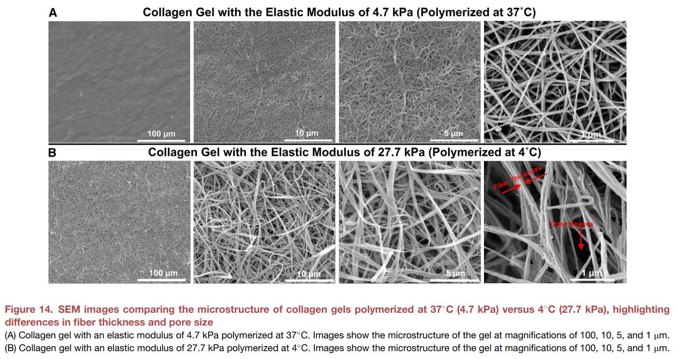
From the publication https://doi.org/10.1016/j.matt.2025.102094, we see that telocollagen hydrogels polymerized at 37C form thinner fibers, with decreased porosity, and softer hydrogels.
Polymerizing telocollagen at 4C forms thicker fibers, increased porosity and firmer hydrogels. The stiffness values were evaluated by individual fibers, not bulk hydrogel stiffness.
Product Applications
Protocol: Cell Seeding & Encapsulation in Collagen
Protocol: Hydrogel Dehydration for SEM
Protocol: Hydrogel Embedding
Protocol: Live Dead Cell Viability
Protocol: Immunofluorescence Staining
Protocol: RNA Extraction for Cells in Hydrogel
Protocol: Actin Cytoskeleton Staining
Protocol: Stem Cell Culture & Differentiation
Protocol: Hydrogel H&E Staining
Protocol: AlamarBlue Cell Proliferation Assay on PureCol
Read our Collagen Hydrogel Shear Modulus whitepapers
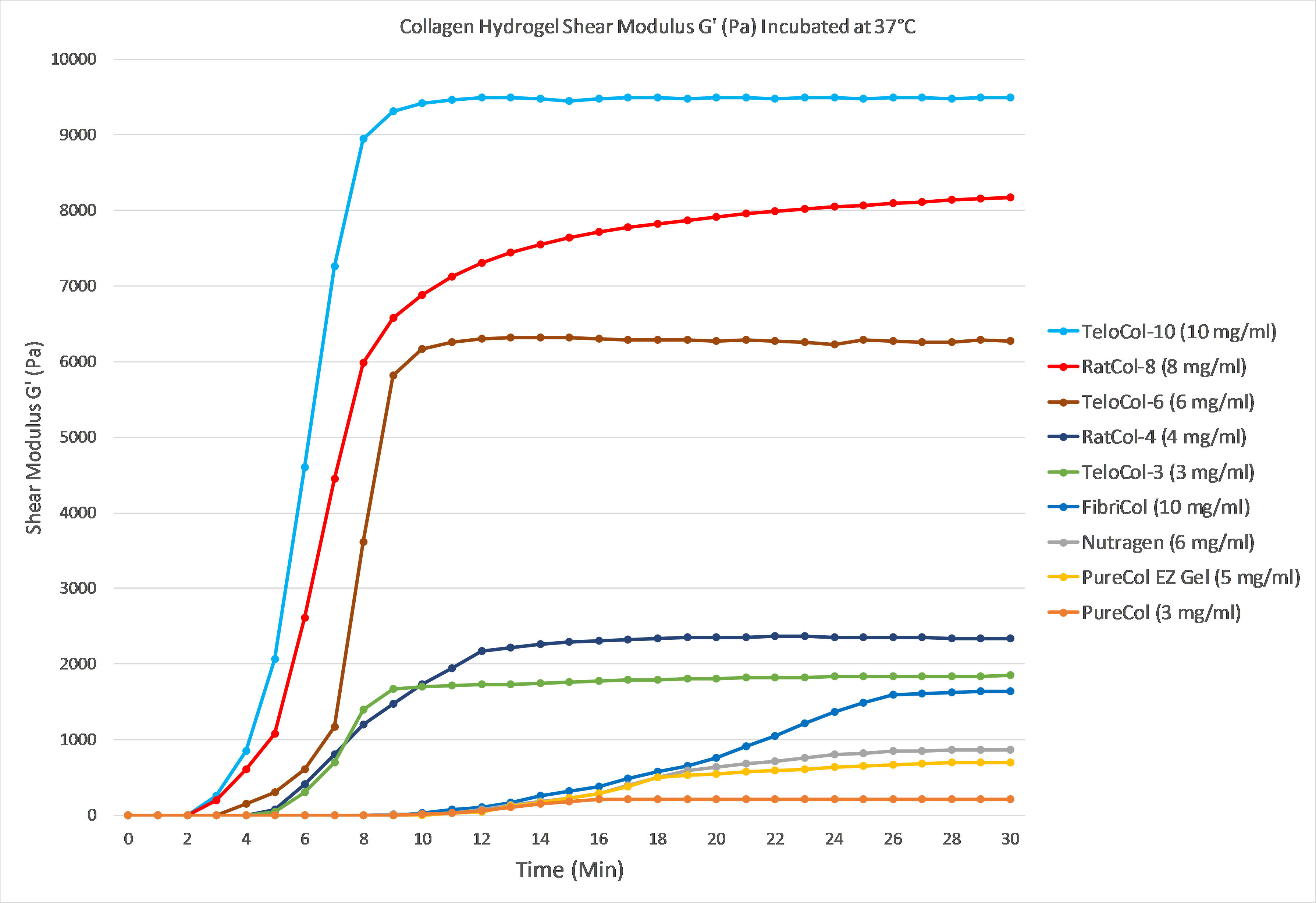
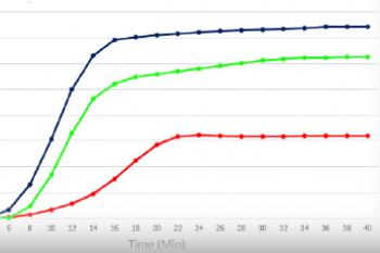
The graph below demonstrates gelation kinetics of our collagen hydrogels. The collagen was neutralized according to respective protocols and placed in specialized containers within the 37°C chamber of the rheometer. Shear Modulus G' (Pa) is shown on the Y axis compared to Time (minutes) on the X axis. All of the below products are in inventory and are available for order.
Product References
References for RatCol®:
Dollinger, Bryan R., et al. "Reactive oxygen species shielding hydrogel for the delivery of adherent and nonadherent therapeutic cell types." Tissue Engineering Part A 23.19-20 (2017): 1120-1131.
Jia, Hong, et al. "The tumor cell‐secreted matricellular protein WISP1 drives pro‐metastatic collagen linearization." The EMBO Journal (2019).
Pérez Cardona, David José. "Phenotype analysis of dermal-epidermal organotypics made with hacat cell line seeding on top of three human fibroblast populated matrices (polyethylene terephthalate, fibrin or collagen I+ III)." (2017).
Karki, Surya B., et al. "Investigation of non-thermal plasma effects on lung cancer cells within 3D collagen matrices." Journal of Physics D: Applied Physics 50.31 (2017): 315401.
Keating, M., et al. "Spatial distributions of pericellular stiffness in natural extracellular matrices are dependent on cell-mediated proteolysis and contractility." Acta biomaterialia 57 (2017): 304-312.
Blum, Kevin M., et al. "Acellular and cellular high-density, collagen-fibril constructs with suprafibrillar organization." Biomaterials science 4.4 (2016): 711-723.
Kaufman, Gili, and Drago Skrtic. "Spatial development of gingival fibroblasts and dental pulp cells: Effect of extracellular matrix." Tissue and Cell 49.3 (2017): 401-409.
Product Certificate of Analysis
Product Videos

Product Disclaimer
This product is for R&D use only and is not intended for human or other uses. Please consult the Material Safety Data Sheet for information regarding hazards and safe handling practices.