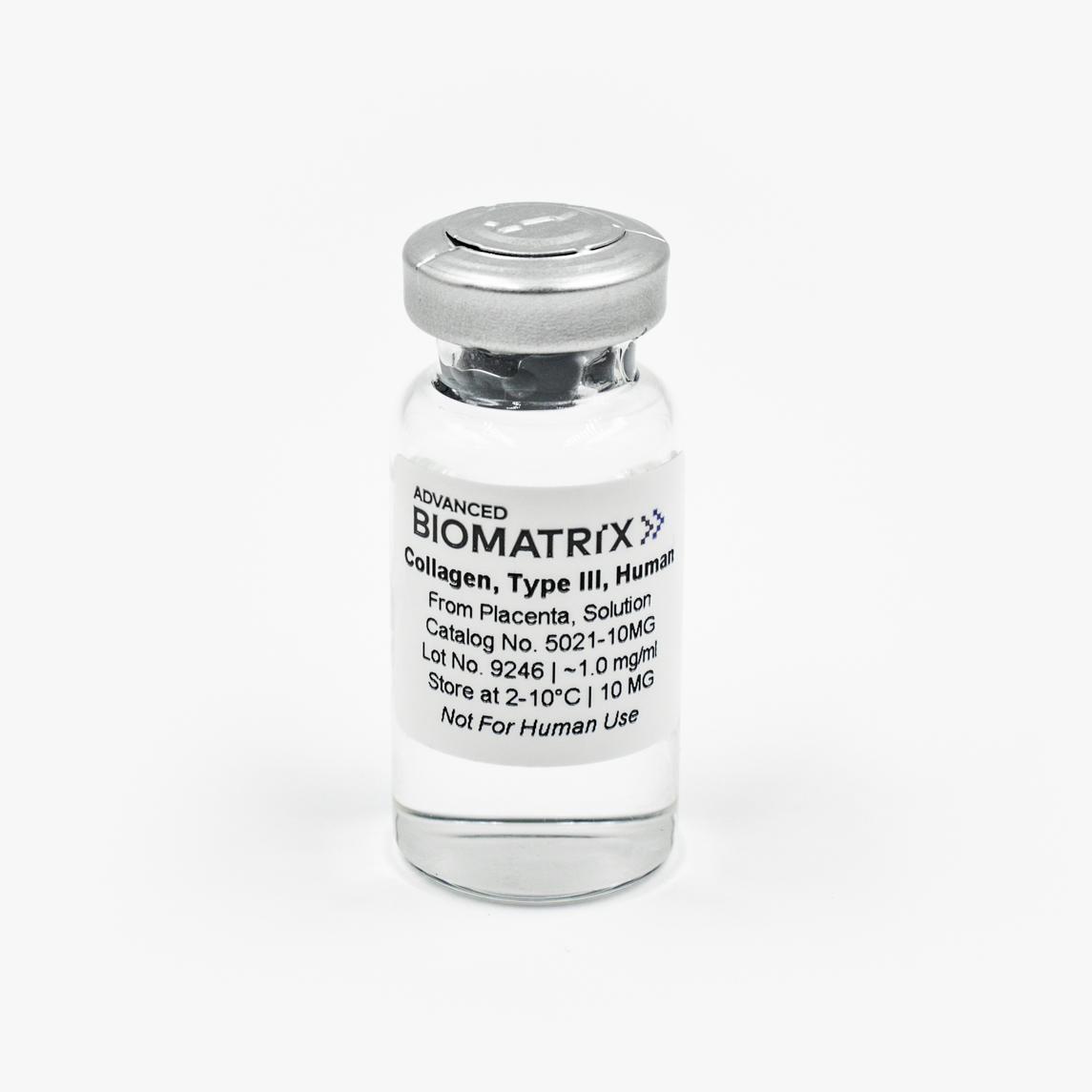
Type III Collagen
Solution, 1 mg/ml (Human)
Catalog #5021
Type III Collagen
Solution, 1 mg/ml (Human)
Catalog #5021
This Type III Collagen product is isolated from human placenta and is purified using a multi-step process with approximately 85% Type III collagen with the remainder being comprised of Type I collagen. The product is supplied as a sterile solution with 10 mg at approximately 1 mg/ml in 0.01 N HCl, pH 2.
Product Description
This Type III Collagen product is isolated from human placenta and is purified using a multi-step process with approximately 85% Type III collagen with the remainder being comprised of Type I collagen. The product is supplied as a sterile solution with 10 mg at approximately 1 mg/ml in 0.01 N HCl, pH 2. Type III collagen product is provided in a user-friendly packaging for use and storage. This Type III collagen product is sterile filtered and is supplied as a ready to use solution.
Type III Collagen provides structure and strength to connective tissue, is found many places in the body, especially skin, lung, intestinal walls and the walls of blood vessels. Collagen III is initially produced as procollagen, a protein consisting of three pro-alpha1(III) chains that form the triple-stranded, rope-like molecule. After being synthesized, the procollagen molecule is modified by the cell. Enzymes modify the amino acids lysine and proline in the protein strands by adding chemical groups that are necessary for the strands to form a stable molecule and then later to crosslink to other molecules outside the cell. The Type III procollagen molecules are released from the cell and are processed by enzymes that clip small segments off either end of the molecules to form mature collagen. The mature collagen molecules assemble into fibrils. Cross-linking between molecules produces a very stable fibril, contributing to collagen’s tissue strengthening function.
Type III Collagen is typically used as a thin coating on tissue culture surfaces or as a control. Specific instructions are found in the Directions for Use. This product is generally used in vitro as a substrate scaffold to enhance cell attachment, adherence and proliferation. Type III collagen may be used to culture a variety of cell types.
| Parameter, Testing, and Method | Type III Collagen #5021 |
| Sterilization Method | Filtration |
| Form | Solution |
| Package Size | 10 mg (~10 mL) |
| Storage Temperature | 2-10°C |
| Shelf Life | Minimum of 6 months from date of receipt |
|
Collagen Concentration - Biuret |
0.7-1.25 mg/mL |
| pH | 1.8-2.5 |
|
Adventitious Agents - by RPR |
Human source has been tested and found negative for Hepatitis B, C, Syphilis and immunodeficiency virus-1. |
|
Electrophoretic Pattern - Coomassie Blue |
Characteristic |
| Sterility - USP modified | No growth |
| Endotoxin (LAL) | <10.0 EU/mL |
| Osmolality (mOsmo H2O/kg) | <35 |
| Cell Attachment Assay | Pass |
| Source | Human Placenta |
Directions for Use
Recommended Volumes for 2D Coatings or 3D Hydrogels
Download the full PDF version or continue reading below:
Coating Procedure
Note: Use these recommendations as guidelines to determine the optimal coating conditions for your culture system.
- Remove required quantity of collagen from the bottle and dispense into a dilution vessel.
- Dilute Type III collagen solution with a 0.01 M HCl solution water to ~50 to 100 µg/ml (~1:10).
- Swirl contents gently until material is completely mixed.
- Add appropriate amount of diluted Type III Collagen to the culture surface ensuring that the entire surface is coated.
- Incubate at room temperature, covered for 1-2 hours.
- After incubation, aspirate any remaining material.
- Rinse coated surfaces carefully with sterile medium or PBS, avoid scratching surfaces.
- Coated surfaces are ready for use. They may also be stored at 2-8°C damp or air dried if sterility is maintained.
Product Q & A
The Type III collagen undergoes an enzyme step in the purification process, resulting in Atelocollagen.
The purity is determined using an interrupted gel electrophoresis as seen in this report:
We have solubilized the Type III collagen at 10 mg/ml, though it is a very viscous solution.
Product References
References for Type III Collagen:
Drzewiecki, Kathryn E., et al. "Circular dichroism spectroscopy of collagen fibrillogenesis: a new use for an old technique." Biophysical journal 111.11 (2016): 2377-2386.
Cho, Soo Hyun, et al. "Meat quality traits as a function of cow maturity." Animal Science Journal 88.5 (2017): 781-789.
Da, Qi, et al. "Fluorescent labeling of endogenous platelets for intravital microscopy: Effects on platelet function." Microcirculation 25.6 (2018): e12457.
Jerrell, Rachel J., Mitchell J. Leih, and Aron Parekh. "The altered mechanical phenotype of fetal fibroblasts hinders myofibroblast differentiation." Wound Repair and Regeneration 27.1 (2019): 29-38.
Simmons, Chad. "The Effect of 3D Collagen Scaffolds on Regulating Cellular Responses." (2015).
Product Certificate of Analysis


Product Disclaimer
This product is for R&D use only and is not intended for human or other uses. Please consult the Material Safety Data Sheet for information regarding hazards and safe handling practices.