Hydrogel H&E Staining Protocol
08/27/24
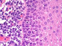
Hematoxylin and eosin (H&E) staining is a widely used technique in histology and pathology for the visualization of tissue architecture.
Hematoxylin stains cell nuclei blue or purple, while eosin stains the cytoplasm and extracellular matrix pink or red.
This contrast allows for detailed examination of tissue morphology, making H&E staining an essential tool for diagnosing diseases, studying tissue structure, and conducting research in various biomedical fields.